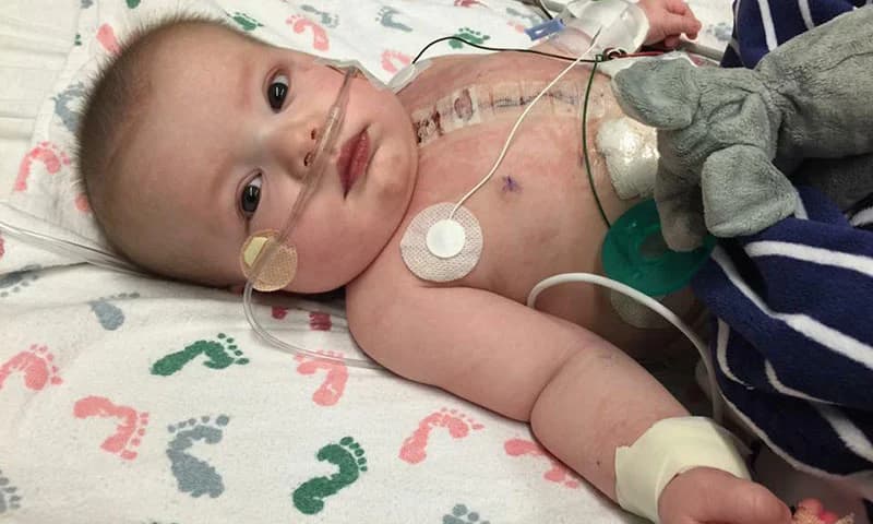
A healthy heart has 4 chambers, a left atrium, and right atrium, and a left ventricle and right ventricle. When a baby is born with a single ventricle defect it means only one ventricle works well enough to pump blood.
What Are the Signs & Symptoms of a Single Ventricle Defect?
A newborn with a single ventricle defect can have:
trouble breathing
trouble feeding
blue or grayish color of the skin and nails
lethargy (very little activity)
weak pulses in the arms and legs
few wet diapers
Your newborn may have the signs a few hours to a day or so after birth. If left untreated, the baby’s blood pressure will fall too low to meet the body's needs, but doctors usually find this issue before birth.

How Is a Single Ventricle Defect Treated?
To repair many types of single ventricle heart defects, including hypoplastic left heart syndrome (HLHS), tricuspid atresia, double outlet left ventricle (DOLV), some heterotaxy defects, and other congenital heart defects, a series of open heart procedures (2 to 3) are performed over several years. This is called “staged reconstruction”. During these procedures, surgeons reconfigure the heart and circulatory systems. Afterward, the heart functions like a one-sided pump with two chambers. The heart no longer receives deoxygenated blood from the veins and this blood flows directly to the lungs instead. As the heart receives oxygenated blood from the lungs, it can then pump it to the body. This is called Fontan circulation. The surgery includes:
Norwood Procedure
Glenn Procedure
Fontan Procedure
The survival rate for children at age 5 is about 70 percent and most of these children have normal growth and development.
Norwood Procedure
Performed shortly after birth, this procedure converts the right ventricle into the main ventricle pumping blood to both the lungs and the body. The main pulmonary artery and the aorta are connected and the main pulmonary artery is cut off from the two branching pulmonary arteries that direct blood to each side of the lungs. A connection called a shunt is placed between the pulmonary arteries and the aorta to supply blood to the lungs instead. Norwood procedure is a type of open-heart surgery for babies born with hypoplastic left heart syndrome, which is usually performed in the first few weeks of life. You should remember, that children with hypoplastic left heart syndrome will need at least two more surgeries, the Glenn procedure (around age 3–6 months) and the Fontan procedure (around age 18–36 months).

What Happens During the Norwood Procedure?
Building a new, larger aorta: The bottom part of the pulmonary artery (which normally goes from the right ventricle to the lungs) is joined with the aorta. This new, bigger aorta now goes from the right ventricle to the body.
Creating a shunt (path) to get blood to the lungs: Because the pulmonary artery now goes from the right ventricle to the body, a shunt is needed to take blood to the lungs. The surgeon will use one of these shunts:
A Blaylock-Taussig-Thomas shunt moves blood from (and through) the aorta to the lungs.
A Sano shunt moves blood from the right ventricle to the pulmonary artery. From the pulmonary artery, the blood goes into the lungs.
Closing the patent ductus arteriosus (PDA): Until now, the baby needed the PDA to stay open so blood could flow from the right ventricle to the body. Now that the right ventricle can pump more blood through the new, bigger aorta, the PDA isn't needed and can be closed.
Making the atrial septal defect (ASD) bigger: This lets more blood with oxygen get back to the right ventricle so it can be pumped out to the body.
Recovery after Norwood Procedure
Your baby will need to spend 3-4 weeks in the hospital to fully recover from the Norwood procedure.
Glenn Procedure
Performed 6 months after the Norwood, Glenn procedure diverts half of the blood to the lungs when circulation through the lungs no longer needs as much pressure from the ventricle. During this procedure, the shunt to the pulmonary arteries is disconnected and the right pulmonary artery is connected directly to the superior vena cava, the vein that brings deoxygenated blood from the upper part of the body to the heart. This sends half of the deoxygenated blood directly to the lungs without going through the ventricle. The Glenn procedure is a type of open-heart surgery Babies who need this surgery typically have it when they’re 4–6 months old.
Why Is the Glenn Procedure Done?
The Glenn procedure is performed for babies born with heart problems like hypoplastic left heart syndrome (HLHS), tricuspid atresia, and double outlet right ventricle. Based on the type of heart problem, your baby may need the Norwood procedure before the Glenn surgery.
What Happens During the Glenn Procedure?
During this surgery, the surgeon disconnects the superior vena cava (SVC) from the heart and connects it to the pulmonary artery. They are allowing blood from the upper part of the body to flow directly into the pulmonary artery. The pulmonary artery then takes the blood to the lungs.
Recovery after Glenn Procedure
Your baby will need to spend 1-2 weeks in the hospital to recover from the Glenn procedure. Depending on the heart problem, a child might need another surgery, the Fontan procedure, when they’re around 18–36 months old. Afterward, your baby will need to see a cardiologist regularly and get EKGs, echocardiograms, lab tests, and occasional cardiac catheterizations.
Fontan Procedure
The third stage of the “staged reconstruction” is performed 18-36 months after the Glenn procedure. Connecting the inferior vena cava, the blood vessel that drains deoxygenated blood from the lower part of the body into the heart, to the pulmonary artery by creating a channel through or just outside the heart to direct blood to the pulmonary artery. At Fontan Procedure, all deoxygenated blood flows passively through the lungs.
Why Is the Fontan Procedure Done?
The Fontan procedure is performed for babies born with heart problems like hypoplastic left heart syndrome (HLHS), tricuspid atresia, and double outlet right ventricle. Based on the type of heart problem, your baby may need the Norwood procedure and Glenn procedure before the Fontan surgery.
What Happens During the Fontan Procedure?
I was disconnecting the inferior vena cava (IVC) from the heart and connecting it to the pulmonary artery using a conduit (tube).
Making a small hole between the conduit and the right atrium, which allows some blood to flow back to the heart. It prevents too much blood from flowing to the lungs right away so they have time to adjust. Doctors can close the fenestration later by doing a cardiac catheterization procedure.
Recovery after Fontan Procedure
Your baby will need to stay in the hospital for 1-2 weeks to recover from the Fontan procedure.
Aftercare for the procedures
Your baby will be getting around-the-clock care and monitoring. Some medication will be given to your baby to help the heart and improve blood flow. Some medication will be needed at home as well. You don’t need to worry, as the care team will teach parents how to care for their baby at home. Also, you can take your baby home, when they are feeding well, growing well, and gaining weight. Afterward, your baby will need to see a cardiologist regularly and get EKGs, echocardiograms, lab tests, and occasional cardiac catheterization.
How Can Parents Help?
Giving any medicines
Feeding your baby
Checking your baby’s weight
Checking oxygen levels with a pulse oximeter
Going to follow-up doctor visits
When Should I Call the Doctor?
You should call the care team immediately if your baby:
Is not eating
Is vomiting
Seems to be breathing fast or struggling to breath
Has oxygen levels that are higher or lower than usual
Seems very irritable
It just doesn't seem quite right
Conclusion
In conclusion, staged reconstruction heart surgery is a life-saving intervention for children born with single ventricle heart defects. This complex process involves a series of surgeries over several years, including the Norwood procedure, Glenn or Hemi-Fontan procedure, and the Fontan procedure. These surgeries reconfigure the heart and circulatory system, allowing the heart to function like a one-sided pump with two chambers. The goal is to improve blood flow and oxygenation, enhancing the child’s quality of life. However, it’s important to note that this treatment requires frequent surveillance in infancy and early childhood to minimize risk factors. Despite the challenges, staged reconstruction heart surgery represents a significant advancement in the treatment of congenital heart defects, offering hope to many families.
Read more: