Cancer diagnosis is a crucial and multifaceted process that aims to detect the presence, type, and extent of cancer in the body. Early and accurate diagnosis is vital for determining the most effective treatment plan and improving the chances of successful outcomes.
Cancer Diagnosis
Cancer diagnosis involves a series of tests and procedures designed to detect and accurately characterize the presence of cancer in the body. The journey typically begins with a thorough medical history and physical examination conducted by a healthcare professional. During this initial assessment, the doctor will evaluate the patient's symptoms and look for signs that may indicate cancer, such as unexplained weight loss, persistent pain, lumps, or changes in bodily functions.
Cancer diagnosis tests
Cancer diagnosis involves a series of sophisticated tests and procedures aimed at detecting and characterizing cancer in the body. These diagnostic methods provide critical information necessary for determining the best course of treatment.
Laboratory tests are often the first step, analyzing samples of blood, urine, and other body fluids. A blood chemistry test examines the levels of various substances in the blood. Abnormal levels can indicate cancer's presence or give clues about cancer spread. For example, elevated calcium levels may suggest bone metastasis.
A complete blood count (CBC) measures the number of different cells in the blood, such as red blood cells, white blood cells, and platelets. Abnormalities in these counts can be a sign of blood cancers like leukemia or lymphoma.
Cytogenetic analysis involves examining the chromosomes in cells to detect genetic abnormalities that may be associated with certain types of cancer. This test is especially useful in diagnosing leukemia and lymphomas.
Immunophenotyping is used to identify specific types of cancer cells based on the markers they express on their surfaces. This test helps in distinguishing between different types of leukemia and lymphoma, which is essential for choosing the appropriate treatment.
Liquid biopsy is a newer, less invasive test that detects cancer cells or fragments of tumor DNA circulating in the blood. This test can provide information about the presence and type of cancer, as well as monitor treatment response and detect recurrences.
Sputum cytology examines mucus (sputum) coughed up from the lungs under a microscope to look for cancer cells, useful in diagnosing lung cancers.
Tumor marker tests measure the levels of specific proteins or substances produced by cancer cells in the blood. Elevated levels of these markers can indicate the presence of certain cancers, such as prostate-specific antigen (PSA) for prostate cancer or CA-125 for ovarian cancer.
Urinalysis is the analysis of urine, which can reveal substances that indicate cancer in the urinary system, such as blood or abnormal cells.
Urine cytology involves examining urine samples under a microscope to detect cancer cells, often used to diagnose cancers of the urinary tract, including bladder cancer.
Each of these diagnostic tests plays a crucial role in the comprehensive assessment of a patient's health. They help confirm the presence of cancer, identify its type and stage, and provide essential information for developing a personalized treatment plan. Advances in these diagnostic techniques continue to improve early detection, enhance treatment outcomes, and ultimately increase survival rates for cancer patients.
Imaging tests
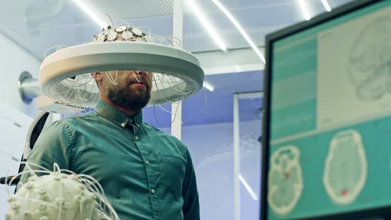
Cancer imaging tests are critical tools in the detection, diagnosis, and management of cancer. These imaging techniques provide detailed visuals of the body's internal structures, helping to identify tumors, assess their size and location, and determine whether cancer has spread.
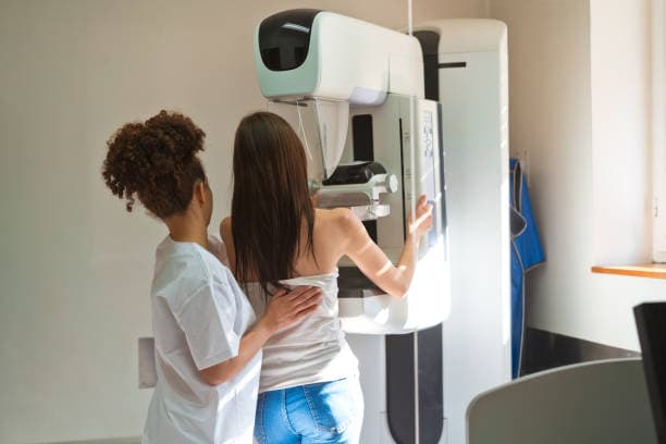
A CT scan (computed tomography) uses X-rays to create cross-sectional images of the body. This imaging method is highly effective for detecting tumors and evaluating their size and exact location. CT scans are often used to guide biopsies and plan treatments, such as surgery or radiation therapy.
Magnetic resonance imaging (MRI) uses strong magnets and radio waves to generate detailed images of soft tissues. This makes it particularly useful for detecting cancers in the brain, spinal cord, and other areas where soft tissue contrast is crucial. MRI scans are excellent for distinguishing between benign and malignant tumors.
Nuclear scans, such as bone scans, involve injecting a small amount of radioactive material into the body. This material accumulates in areas of high metabolic activity, such as cancerous tissue, and is detected by a special camera. Bone scans are specifically used to determine if cancer has spread to the bones, which is common in cancers like breast, prostate, and lung cancer.
Positron emission tomography (PET) scans involve injecting a radioactive sugar substance into the body. Cancer cells, which have higher metabolic rates than normal cells, absorb more of this substance. The PET scanner then detects these areas of high radioactivity, highlighting the presence and spread of cancer. PET scans are often combined with CT scans to provide both metabolic and anatomical information.
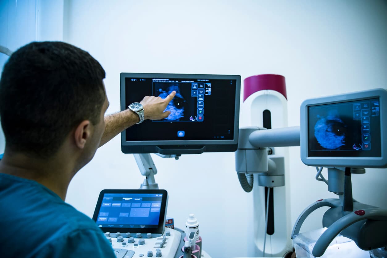
Ultrasound uses high-frequency sound waves to create images of the inside of the body. It is frequently used to guide needle biopsies and to examine organs like the liver, pancreas, and reproductive organs. Ultrasounds are non-invasive and do not involve radiation, making them a safer option for certain patients.
X-rays are one of the oldest and most commonly used imaging techniques. They are particularly useful for examining bones and detecting lung cancers. Despite being less detailed than CT or MRI scans, X-rays are quick and widely available, making them a valuable tool for initial cancer assessments.
Each of these imaging tests has its strengths and is often used in combination to provide a comprehensive view of the cancer's characteristics. The choice of imaging technique depends on various factors, including the type of cancer, its location, and the specific information needed for diagnosis and treatment planning. Advances in imaging technology continue to enhance the precision and effectiveness of these tests, contributing significantly to the fight against cancer.
Cancer diagnosis procedure
A biopsy is a critical cancer diagnosis procedure involving the removal of a small tissue sample from a suspicious area to be examined under a microscope. It is the gold standard for confirming the presence and type of cancer. The process begins with an initial consultation where a healthcare professional assesses the patient's symptoms, medical history, and the results of preliminary tests like imaging studies or lab work. Based on these evaluations, a biopsy may be recommended if there are indications of abnormal growths or suspicious lesions.
There are several types of biopsies, each suited for different situations. The choice of biopsy method depends on the location and accessibility of the suspected tumor. One common type is a needle biopsy, which includes fine-needle aspiration (FNA) and core needle biopsy. FNA uses a thin needle to extract a small amount of tissue or fluid, while a core needle biopsy uses a larger needle to obtain a more substantial tissue sample.
Another method is a surgical biopsy, which can be either incisional or excisional. An incisional biopsy involves removing a portion of the suspicious area, while an excisional biopsy involves removing the entire lesion or tumor. Surgical biopsies are typically performed under local or general anesthesia in an operating room or specialized procedure room. These are often used when the tumor is easily accessible or when a larger tissue sample is needed for a more definitive diagnosis.
Endoscopic biopsy is another technique where a thin, flexible tube with a light and camera (endoscope) is inserted through natural body openings (like the mouth or rectum) or small incisions to reach internal organs. Tools attached to the endoscope can then be used to collect tissue samples. This method is frequently used for cancers of the digestive tract, lungs, and other internal organs.
The collected tissue samples are sent to a pathology lab, where a pathologist examines them under a microscope to identify cancer cells. The pathologist's report provides crucial information about the type, grade, and stage of cancer, which helps guide treatment decisions.
What happens if there's cancer in your scans?
If your scans reveal the presence of cancer, it's natural to feel a mix of emotions, from fear and uncertainty to determination and hope. The next steps are crucial in understanding your condition and initiating the best possible treatment plan.
Firstly, your healthcare provider will likely refer you to an oncologist, a specialist in cancer treatment. This expert will conduct a comprehensive evaluation, which may include additional tests such as blood work, biopsies, and advanced imaging to confirm the diagnosis and understand the extent (stage) of the cancer.
Upon confirmation of the diagnosis, your oncologist will discuss the type and stage of cancer with you, providing a detailed explanation of what this means for your health and treatment options. It’s important to ask questions and seek clarity on any concerns you may have. Understanding your diagnosis empowers you to make informed decisions about your care.
Next, a personalized treatment plan will be developed based on the type, stage, and location of the cancer, as well as your overall health and preferences. Treatment options may include surgery to remove the tumor, radiation therapy to target cancer cells, chemotherapy to kill or slow the growth of cancer cells, targeted therapy that attacks specific cancer cell mechanisms, immunotherapy to boost the body's natural defenses, and other advanced treatments like hormone therapy or clinical trials.
Throughout this process, it’s essential to have a strong support system. Family, friends, and support groups can provide emotional encouragement and practical assistance. Additionally, consider seeking a second opinion from another oncologist or cancer center to ensure that you are exploring all available options.
Maintaining open communication with your healthcare team is key. Keep track of your symptoms, side effects, and any changes in your condition, and share this information during your appointments. Adhering to your treatment plan and attending regular follow-up visits are vital for monitoring your progress and making necessary adjustments to your care.
Lifestyle changes can also play a significant role in your overall well-being. A balanced diet, regular physical activity, stress management techniques, and adequate rest can help support your body during treatment and recovery. If you smoke, quitting can improve your prognosis and treatment outcomes.
Navigating a cancer diagnosis is undoubtedly challenging, but staying informed, proactive, and connected with your healthcare team can make a significant difference in your journey. Advances in cancer research and treatment continually offer new hope, and many patients successfully manage their condition and lead fulfilling lives.
Conclusion
In conclusion, cancer diagnosis is a multifaceted and essential process that combines advanced medical techniques and thorough assessments to detect, characterize, and understand cancer. Early and accurate diagnosis is crucial for determining the most effective treatment plan and improving patient outcomes. By employing a variety of diagnostic tools, such as imaging studies, laboratory tests, and biopsies, healthcare professionals can gather comprehensive information about the type, stage, and spread of cancer. This information guides personalized treatment plans, enabling targeted therapies and improving the chances of successful treatment. While a cancer diagnosis can be daunting, advances in medical science, coupled with a strong support system and proactive healthcare management, provide hope and pathways to recovery for many individuals facing this challenging journey.
Read More